Hello everyone, Dr. Wan here.
Let’s talk about ears. Specifically, let’s talk about when ears decide to go rogue.
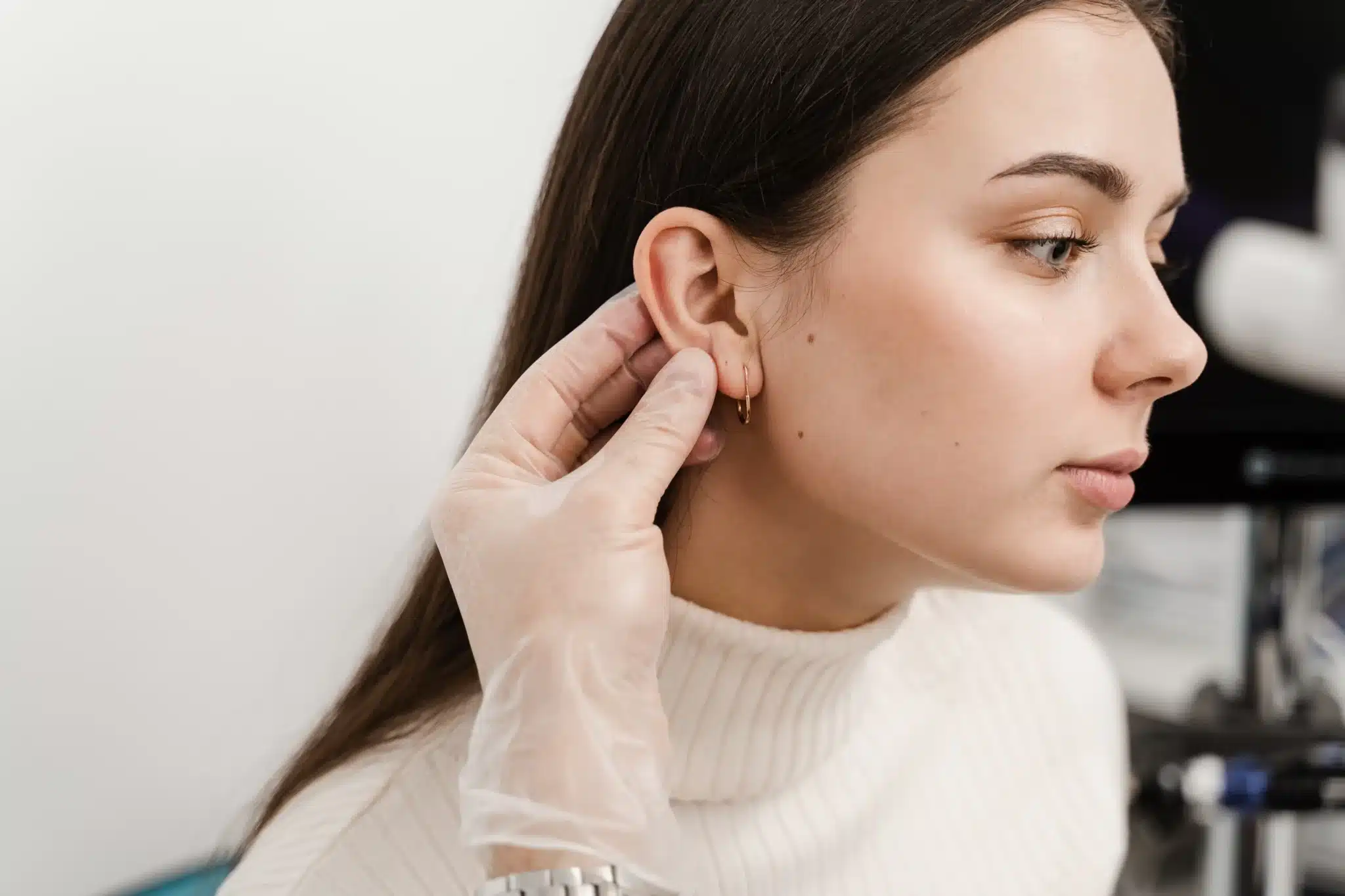
If you’re reading this, there’s a good chance you or someone you know is dealing with an ear keloid. You know the story: it started as a fun piercing—maybe a helix, maybe a simple lobe—and then, months later, a small bump appeared. You ignored it. It got bigger. You panicked. Maybe you went to a GP, got a standard injection, it shrank, and then… boom. It came back, angrier and itchier than before.
I see this frustration every single day. Patients often come to us feeling like they’ve been fighting a losing battle, having tried creams, clips, and single-drug injections that just didn't stick. We often discuss the role of genetics in these cases (you can read more about that in our article Are You Genetically Prone to Keloids? Here's How to Know), but regardless of your DNA, the history of treating these stubborn bumps is a fascinating journey of trial, error, and eventually, scientific triumph.
We’ve moved from the brute force of the scalpel to what I like to call "biological warfare"—outsmarting the cells that cause the scar.
Today, I want to take you through the history of ear keloid management. We’re going to look at why the old ways failed, why some clinics still use them (unfortunately), and how the field has arrived at the current state of the art—a comprehensive management philosophy we are very passionate about here at 1Aesthetics.
The Antagonist: Why Your Ear is Being Difficult
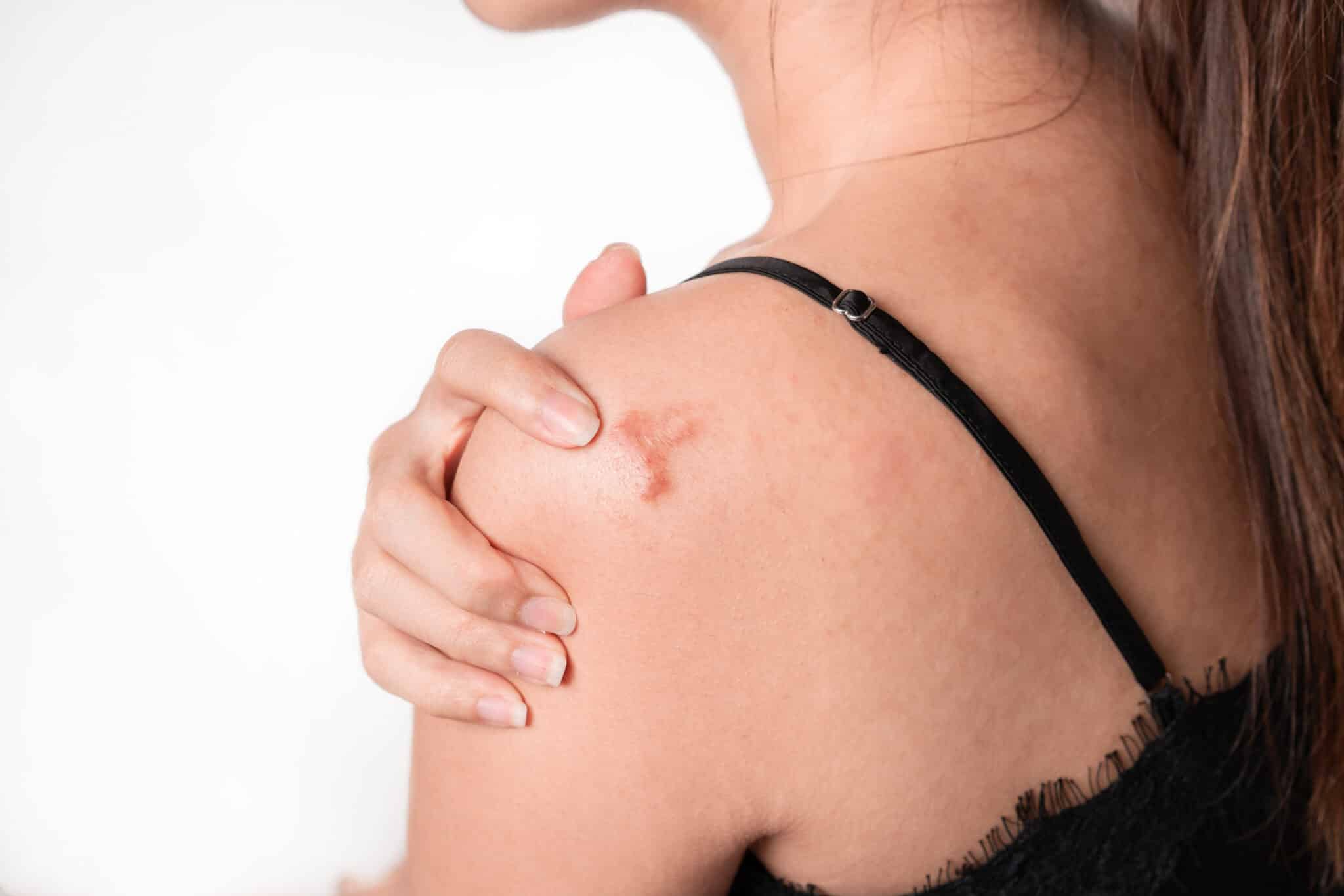
Before we dive into the history books, we have to understand the enemy. A keloid isn't just a scar; it’s a biological error.
For a long time, doctors believed keloids formed purely because of "high tension" (skin being stretched tight). But if you think about it, the earlobe is actually an area of minimal skin tension compared to, say, your chest or shoulder. Yet, it is one of the most common sites for keloids.
So, what gives?
Recent research points to something more complex. The ear is unique because the skin is splinted by the underlying cartilage. When you pierce through the "transitional zone" (where the cartilage meets the soft tissue) or the cartilage itself, you aren't just injuring skin; you are triggering a reaction in the cartilage cells (chondrocytes) and upregulating specific proteins like COMP (Cartilage Oligomeric Matrix Protein). This creates a unique inflammatory storm. The cartilage acts as a rigid backboard, and when the inflammation hits, the fibroblasts (scar-forming cells) go into overdrive, pouring "cement" (collagen) 24/7. This is an inflammatory and cellular issue—not just a "tight skin" issue—is why modern management has shifted from simple cutting to biological modulation. For a broader overview of how we approach this, check out our page on Keloid Scar Removal Treatment in Singapore.
The "Dark Ages": A History of Surgical Failures
In the early days of dermatology and plastic surgery, the approach was intuitive but biologically flawed. The logic was: "There is a lump. Cut it off."
- The "Iceberg" Mistake: Shave Excision Years ago, a common technique was the "shave excision." A provider would take a blade and slice the keloid flush with the skin, like shaving off a mole.
- The Logic: It’s quick, easy, and requires no stitches.
- The Reality: This is arguably the worst thing you can do to a keloid. You’ve only removed the visible tip of the iceberg. The "roots" (the reticular vascular network) are still deep in the dermis. Worse, you’ve left a raw, open wound that has to heal from the bottom up. This causes massive inflammation. The keloid almost always returns, usually twice as fast and twice as wide. The Recurrence Trap: Simple Excision** Realizing that shaving didn't work, surgeons started cutting the whole thing out and stitching the skin together.
- The Logic: If we remove the roots, it won't grow back.
- The Reality: While better than shaving, historical data shows that simple excision alone has a recurrence rate of anywhere from 45% to 100%. By creating a new surgical wound, we often just restart the inflammatory cycle.
- The "High-Tech" Trap: Laser Vaporization I have to mention this because patients ask me about it constantly. "Dr. Wan, can't you just laser it off?"
- The Reality: For a big, bulky keloid? No. Lasers work by burning tissue (thermal ablation). If you try to burn down a thick keloid, you are creating a deep burn wound with a lot of dead, charred tissue (necrosis). This causes profound inflammation. It’s like trying to put out a fire with gasoline. While we love lasers for polishing a flat scar later on (such as with our Fractional CO2 Laser), using them to remove the bulk is a recipe for disaster.
Why Do Some Clinics Still Use These Methods?
You might be wondering: If the medical literature shows these techniques have high failure rates, why are they still being offered?
It’s rarely out of malice. It’s usually a combination of three factors:
- The "Mole" Mindset: Many practitioners spend 99% of their time removing benign moles, cysts, or skin tags. The standard protocol for those is "shave it or cut it." If they don't specialize in scar pathology, they may apply that same logic to a keloid. But a keloid isn't a passive lump; it's a rebellion. Treating it like a mole is the first mistake.
- The "Magic Wand" Fallacy: Lasers are powerful marketing tools. It is much easier to sell a "quick laser removal" than a complex surgical flap reconstruction. Patients often demand lasers because they perceive them as high-tech and scar-less, and clinics may feel pressured to provide what the patient asks for, even if it's the wrong tool for the job.
- The Follow-Up Gap: This is the biggest one. A surgeon might cut off a keloid beautifully and send the patient home. If they don't have a rigorous long-term follow-up protocol, they assume the surgery was a success. But keloids play the long game—they return 6, 12, even 18 months later. By then, the patient has often lost faith in the original doctor and moved on to a specialist (like us) to fix the recurrence. The original doctor never learns that their technique failed, so they keep doing it.
The "Over-Correction": Grafts, Flaps, and Wedges
When simple cutting failed, the medical community swung the other way, trying complex reconstruction techniques borrowed from cancer surgery. While these have their place, they are often overkill for keloids.
- The "Patchwork" Problem: Skin Grafts Some surgeons try to cover the wound with a skin graft—taking a piece of skin from behind the ear, the neck, or the thigh and stitching it onto the ear.
- The Issue: A graft is a piece of skin completely detached from its blood supply. It relies on the wound bed to survive. If it fails, you have a necrotic mess. Even if it survives, the color and texture rarely match the ear perfectly, leaving a "patchwork quilt" appearance. Plus, you now have a second wound at the donor site that could potentially turn into a keloid itself. The "Collateral Damage": Complex Flaps & Undermining** To reduce tension, some techniques involve "undermining" (cutting under the healthy skin to loosen it) or rotating large flaps of tissue from the neck or scalp.
- The Issue: While this reduces tension, it involves extensive cutting of healthy tissue. In a patient whose body loves to make keloids, making the surgical field larger is risky. It’s like poking the bear. We want to minimize trauma, not spread it around.
- The "Deformity": Wedge Resection. This involves cutting out a pie-shaped slice of the ear (cartilage and all) and stitching the edges together.
- The Issue: It works to remove the mass, but it often changes the shape of the ear, leaving it notched, cupped, or smaller than the other side. We believe you shouldn't have to trade a scar for a deformity.
The Turning Point: Respecting the Tissue
The evolution of management finally took a turn for the better when the medical community stopped trying to fight the ear and started working with it. We realized that the skin stretching over the keloid wasn't diseased—it was actually useful. The keloid, in its slow growth, had acted as a natural tissue expander.
This led to the development of the Core Fillet concept (also known as Intralesional Excision).
Think of the keloid like a fruit—say, a lychee. The old way was to throw the whole lychee away. The modern way is to carefully peel the skin (the rind), remove the seed (the hard collagen core), and then lay the skin back down.
Because the skin is preserved, there is plenty of fabric to cover the wound. It can be closed with zero tension. By removing the tension and preserving the anatomy, we remove a primary trigger for regrowth while keeping the ear looking like an ear.
The Modern Dilemma: To Radiate or Not to Radiate?
So, the core is gone. The ear looks great. Are we done? Absolutely not.
This is where the "State of the Art" really comes into play. Removing the bulk removes the product of the disease, but it doesn't cure the process. The fibroblasts are still there, and they are waking up, ready to riot. We need an adjuvant (an assistant) to keep them asleep.
In many hospitals, the "Gold Standard" is listed as Surgery + Immediate Radiotherapy. They blast the ear with radiation to kill the dividing cells [11].
Does it work? Yes. Do we like it for our patients in Singapore? Generally, no.
Here is the 1Aesthetics perspective on why we usually steer clear of radiation for benign ear keloids:
- The "C" Word: Radiation carries a risk—however small—of carcinogenesis (causing cancer) in the surrounding healthy tissue or the thyroid. For a benign lump on the ear, taking even a 0.1% risk of cancer seems unnecessary to me when we have other options.
- The "Zebra" Effect: Radiation kills pigment cells. In Asian skin, this often leads to permanent white patches (hypopigmentation) or dark patches. You might trade a keloid for a permanent white spot.
- Logistics: It’s expensive and requires trips to an oncology center.
The 1Aesthetics Protocol: The Multimodal "Chemical Blockade"
If we don't use radiation, what do we use? We use a "chemical blockade." We have evolved a protocol that may rival the success rates of radiation but with a much better safety profile.
We don't rely on a single "magic" drug. We use a Multimodal Cocktail—a synergy of medications that attack the keloid pathway from different angles. We don't believe in a "one-size-fits-all" mix; we tailor the ingredients to how aggressive your scar is.
- The Anti-Inflammatories: These are the foundation. They suppress the acute inflammation that happens right after any procedure. By calming the immune response, we prevent the "red alert" signal from going out to the scar-forming cells [12].
- The Cell-Cycle Inhibitors (Antimetabolites): This is where modern science shines. These agents stop the fibroblasts from replicating their DNA. It essentially shuts down the factory floor. Studies have shown that adding these agents is often superior to using anti-inflammatories alone and can reduce the risk of skin thinning [13].
- The Tension Relievers (Neuromodulators): "Wait, Dr. Wan, isn't that for wrinkles?" Yes, but it’s also a game-changer for scars. By injecting these into the muscles around the ear and the wound edges, we do two things. First, we relax the tiny muscles that pull on the wound. Second, new research suggests that they actually block the chemical signals that tell the body to make scar tissue [14].
- The Structural Modifiers. For particularly stubborn or hard keloids, we have other tools in the arsenal. These include agents that work on the cellular channels (like calcium channels) to disrupt the structure of the collagen itself. By altering the internal environment of the cell, we can keep the tissue soft and prevent it from organizing into a hard lump.
- The Enzymatic Agents Sometimes, we need to break down the "cement" directly. We can employ specialized enzymes that degrade the specific components of the scar tissue matrix. This helps to soften the lesion and allows our other medications to penetrate deeper into the core.
- The Vascular Inhibitors Keloids are hungry; they need a blood supply to grow. We can include agents that specifically target the tiny blood vessels feeding the scar. By cutting off the supply lines, we starve the keloid of the nutrients it needs to expand.
(For a deeper dive into how we mix and match these treatments, read our detailed guide on Keloid Scar Removal Injections - Beyond Just Steroids)
The "Marathon" Mindset
Here is the most important thing I tell my patients: Keloid management is not an event; it is a relationship.
In the old days, a provider might remove the keloid and say, "Good luck!" That is a recipe for recurrence. At 1Aesthetics, our protocol is a marathon.
- Step 1: We ensure the bulk is addressed using current, tissue-preserving concepts available.
- Step 2: We hit the "pause" button on healing with our intraoperative blockade.
- Step 3: We see you. A lot.
We schedule prophylactic serial injections. We don't wait for the keloid to come back. We treat the healing ridge at specific intervals tailored to your body's healing speed. We keep those fibroblasts in a state of suspended animation until the "danger zone" of healing has passed.
Conclusion
The history of ear keloid management has been a bumpy road, paved with good intentions and high recurrence rates. But we have learned from those failures. We’ve learned that you can’t just shave it off, you can’t just burn it, and you can’t just cut it out and hope for the best.
The current state of the art—the approach we champion—is about elegance and biology. It’s about preserving your natural tissue and using a sophisticated blend of medicines to retrain your body’s healing response.
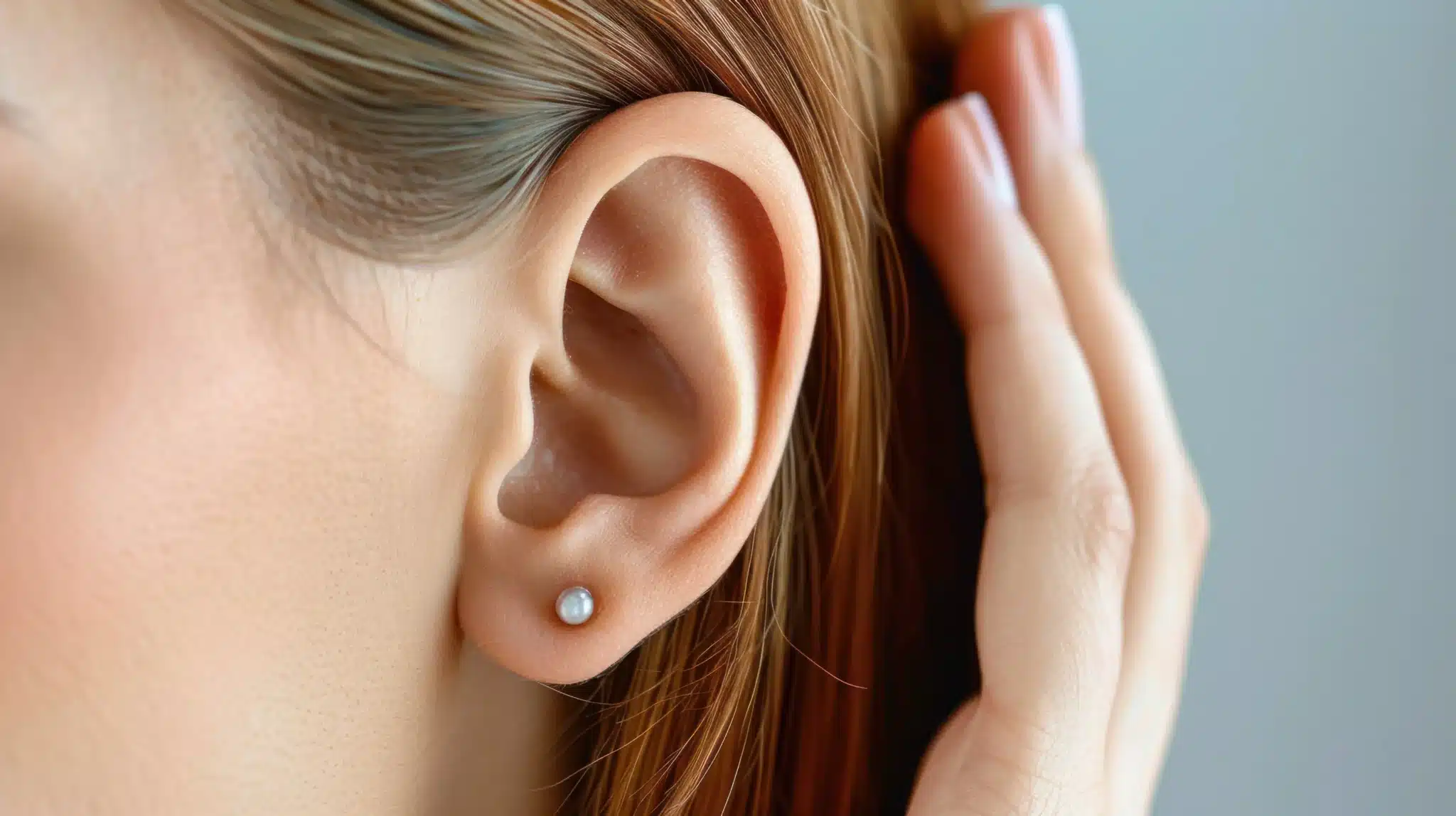
If you are sitting there touching your ear, worrying about that bump, know this: You don't have to live with it, and you don't have to fear the "cut and grow back" cycle. With the right management plan, we can finally get your ears back to being just ears—ready for a nice pair of earrings, or just ready to listen.
Book a Consultation with Dr. Wan | WhatsApp Us
Stay safe, and stop touching that piercing!
Dr. Wan
References
- Ogawa R. The most current algorithms for the treatment and prevention of hypertrophic scars and keloids. Plast Reconstr Surg. 2010;125(2):557-568.
- Zorlu O, Yazici S, Balaban Adım Ş. Keloid formation following ear piercing through the transitional zone. Australas J Dermatol. 2023;64(3):e256-e258.
- Berman B, Maderal A, Steiner A. Keloids and Hypertrophic Scars: Pathophysiology, Classification, and Treatment. Dermatol Surg. 2017;43 Suppl 1:S3-S18.
- O'Sullivan ST, O'Shaughnessy M, O'Connor TP. Ear keloids: a review and update of treatment options. Ir J Med Sci. 2015;184(2):325-330.
- Firoozbakhsh S, et al. Management of Ear Keloids Using Surgical Excision Combined with Postoperative Steroid Injections. World J Plast Surg. 2019;8(3):384-389.
- Leventhal D, Furr M, Reiter D. Treatment of keloids and hypertrophic scars: a meta-analysis and review of the literature. Arch Facial Plast Surg. 2006;8(6):362-368.
- Al-Attar A, Mess S, Thomassen JM, Kauffman CL, Davison SP. Keloid pathogenesis and treatment. Plast Reconstr Surg. 2006;117(1):286-300.
- Kumar A. Piercing Ear Keloid: Excision Using Loupe Magnification and Topical Liquid Silicone Gel as Adjuvant. J Cutan Aesthet Surg. 2017;10(4):204-209.
- Lee Y, Minn KW, Baek RM, Hong JJ. A new surgical treatment of keloid: keloid core excision. Ann Plast Surg. 2001;46(2):135-140.
- Liu CL, Yuan ZY. Retrospective study of immediate postoperative electron radiotherapy for therapy-resistant earlobe keloids. Arch Dermatol Res. 2019;311(6):469-475.
- Danielsen PL, Jorgensen B, Karlsmark T, Jorgensen LN, Agren MS. Triamcinolone acetonide intralesional injection for the treatment of keloid scars: patient selection and perspectives. Patient Prefer Adherence. 2016;10:2181-2188.
- Hietanen KE, Järvinen TA, Huhtala H, Tolonen TT, Kuokkanen HO, Kaartinen IS. Treatment of Keloid Scars With Intralesional Triamcinolone and 5-Fluorouracil Injections - A Randomized Controlled Trial. J Plast Reconstr Aesthet Surg. 2019;72(1):4-11.
- Shaarawy E, Hegazy RA, El-Komy MHM. Botulinum toxin type A: An innovative treatment for hypertrophic scars and keloids. J Cosmet Dermatol. 2021;20(9):2664-2670.