Summary:
When you look at a mole on your face, it may appear no larger than a pencil eraser, but its outward size rarely tells the full story. As the Founder and Medical Director of 1Aesthetics, Medical & Surgery in Singapore, I evaluate moles not just by their surface diameter, but by how deeply their cells are rooted within your skin layers. Two moles that look identical to you can require entirely different surgical or non-surgical approaches, each carrying unique risks and outcomes based on that hidden depth.
In my experience, depth is the critical variable that dictates our treatment path, yet it is often the one factor patients do not consider until we are discussing it together in my consultation room.
There is a common assumption that mole removal is straightforward: remove what you can see, and you are done. In practice, what you see on the skin surface tells only part of the story.
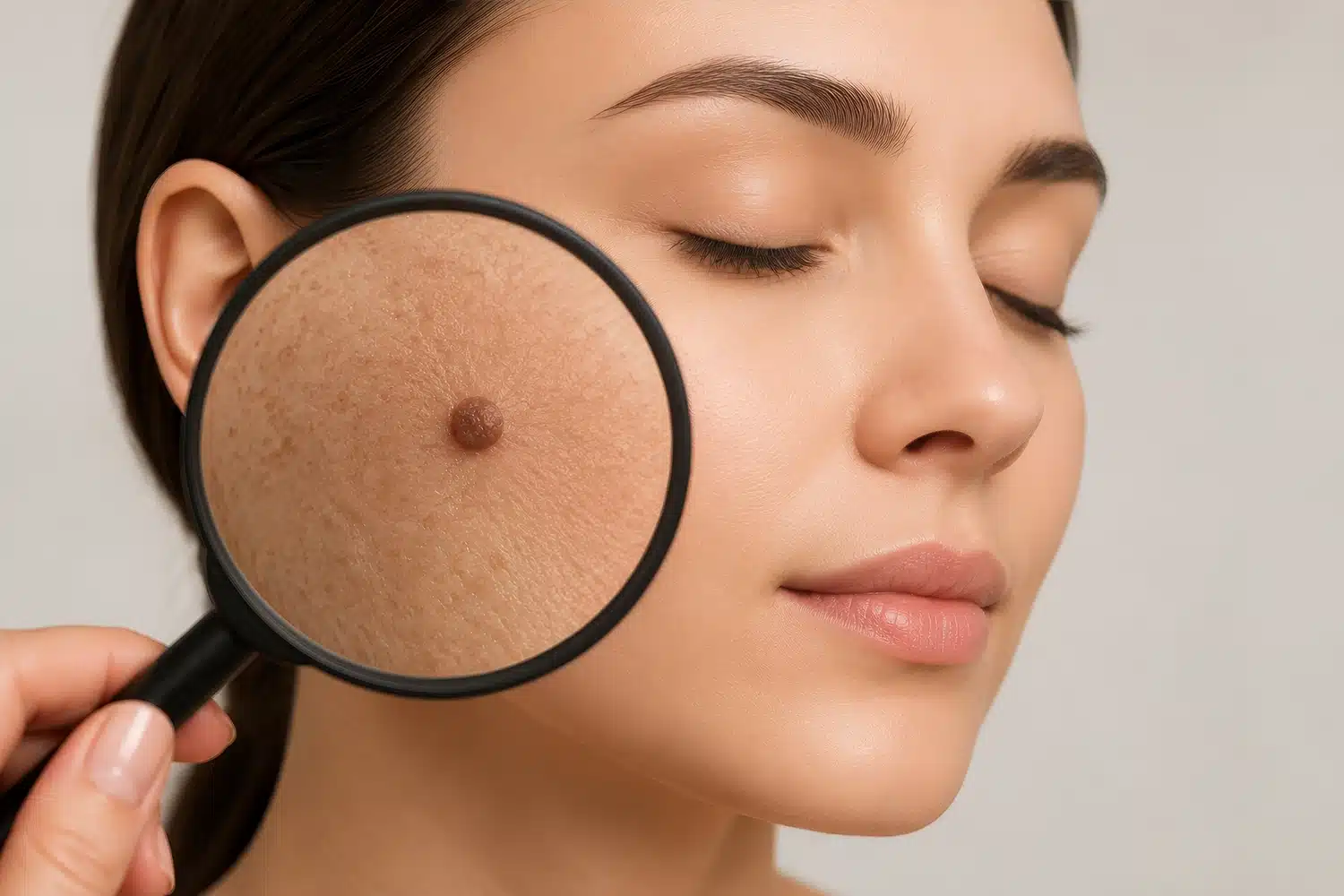
A mole is not simply a flat patch of pigment. It is a cluster of melanocytic cells (pigment-producing cells) that may sit at varying levels within the skin.
One mole might involve only the uppermost layers; another of the same visible diameter might extend well into the dermis (the deeper, structural layer beneath the surface). Size matters, but depth is what determines how complex the removal might be.
A broad, shallow mole can often be addressed by ablative techniques that work through the skin's upper layers without disturbing the underlying tissue. A deeply rooted mole, on the other hand, requires clearance at depth, working through layers of skin that, if disrupted, require careful repair.
The smaller visible footprint of the mole tells you almost nothing about how technically demanding the removal will be.
Moles are typically classified by where their cells are concentrated within the skin. This classification directly informs the treatment plan. The table below shows how the three main types differ and what each typically means for removal.
| Type | Cell location | Typical appearance | Removal implication |
| Junctional naevus | Epidermis/dermis junction | Flat or slightly raised | Generally amenable to non-surgical removal |
| Dermal naevus | Deeper within the dermis | Raised, dome-shaped | Requires reaching deeper tissue; may need excision |
| Compound naevus | Both junction and dermis | Variable | A combination of both considerations above |
Not sure which category your mole falls into? Book a consultation with me at 1Aesthetics to understand your options before deciding anything.
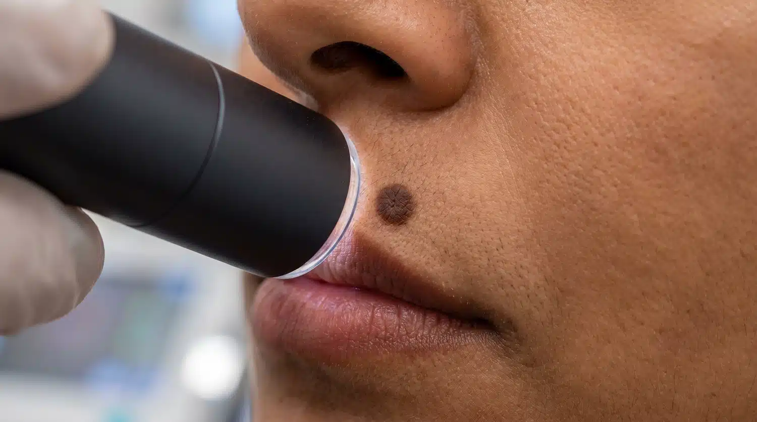
A physician will assess depth using dermoscopy (a handheld magnifying device with polarised light), alongside physical signals such as texture, elevation, and border definition. Where there is uncertainty about the nature of the lesion, a biopsy may be recommended before proceeding.
Blue naevi are a specific type of dermal mole where melanocytes sit unusually deep within the dermis. Their blue or grey appearance is caused by the depth of the pigment beneath the surface.
As with deeply rooted naevi in general, incomplete clearance is the primary risk: if any melanocytic cells are left behind, the mole may recur. There is also a meaningful risk of post-treatment pitting where the depression left by deep tissue disruption does not fill fully during healing.
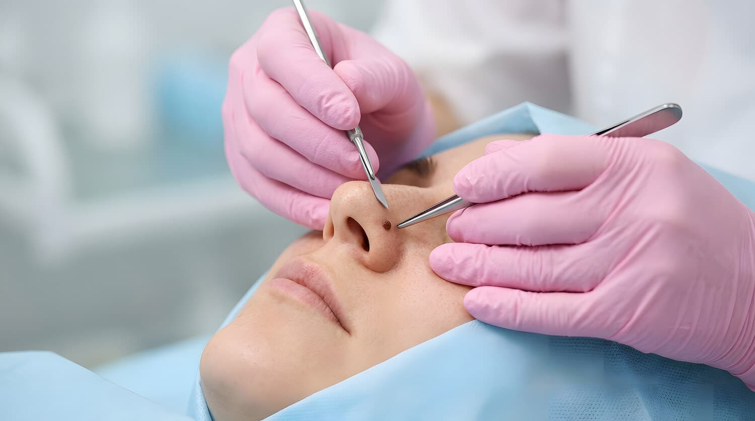
Surgery is not the default for face mole removal in Singapore. The majority of moles on the face can be addressed non-surgically using one of several ablative technologies.
At 1Aesthetics, I explore these four approaches:
For borderline cases where the mole's depth or characteristics make a single non-surgical session insufficient, I may recommend a staged approach across multiple sessions, with growth factors used to support healing in between.
| Treatment | Recommended for | Mechanism |
| Ultrapulse CO2 laser | Raised moles that do not extend deeply | Vaporises tissue layer by layer using water-targeting laser energy |
| Radiofrequency (RF) ablation | Raised moles or those extending slightly deeper | High-frequency energy vaporises or coagulates tissue; insulated needles can reach deeper layers |
| Pigment laser (ruby 694 nm/alexandrite 755nm) | Flat or pigmented moles near the surface | Targets melanin specifically, with less effect on surrounding tissue |
Surgical excision is typically indicated when the mole is deeply rooted, when histological analysis (laboratory examination of tissue) is needed to rule out atypical features, when the mole has recurred following prior treatment, or when the lesion is large enough that ablation alone would not achieve predictable results.
The trade-off is a linear scar in place of the mole, and how visible that scar is depends significantly on where the mole sits and how the wound is closed.
For straightforward cases, I plan each excision to follow the natural lines of the skin where possible, closing the wound with sutures to support healing and reduce scar visibility.
Where the case requires it, I may use other techniques, including:
The natural tension lines of the skin, often referred to as Langer's lines, run in directions that correspond to the underlying orientation of collagen fibres in the dermis.
An incision made parallel to these lines experiences less wound tension during healing and tends to produce a finer, less visible scar. An incision that cuts across them is more likely to widen or thicken as it heals.
Different areas of the face carry different levels of resting tension and movement. The nose and the area around the mouth are among the more technically demanding sites because the tension vectors are complex and the skin is less forgiving of imprecise closure.
At 1Aesthetics, I typically plan the scar direction to sit within or parallel to natural creases where possible.
Where your mole sits matters as much as what it looks like. If you are weighing up different facial mole removal approaches, book an appointment with me so we can discuss whether a surgical approach suits your situation.
In the first five to seven days following mole removal, the skin is in its initial closure phase: keratinocytes (skin cells) migrate across the wound surface, collagen begins to form, and a scab or crust develops.
This is normal and should not be interfered with. The skin's regeneration cycle takes roughly four weeks, and it is at this point that a clearer picture of the healing result begins to emerge.
Scar remodelling is an ongoing process that continues for several months after the wound closes, with gradual changes in texture, colour, and flatness. Sun protection is essential during healing, as UV exposure can stimulate melanin production in the healing skin, leading to post-inflammatory hyperpigmentation (PIH).
I also recommend that you wear protective clothing and consistently use a broad-spectrum sunscreen with an SPF of at least 30.
Healing timeline at a glance:
If you have been thinking about a facial mole and are not sure where to start, the most useful first step is an assessment with a physician who can evaluate the mole directly rather than advise in general terms.
At 1Aesthetics, I assess each case individually, taking into account the mole's depth, location, size, and your skin type before making any treatment recommendation.
Get in touch to find out which mole removal approach is right for your mole.
Address:
1Aesthetics, Medical & Surgery
#14-90 The Central Tower 1
8 Eu Tong Sen Street
Singapore 059818
Mon-Fri 10 AM to 730 PM
Sat 10 AM to 6 PM
Phone / WhatsApp:
+65 66125173 / +65 84899962
Email:
[email protected]