Summary:
Not every mole is what it appears to be, and that is why the first step in any removal decision matters so much. A mole that looks harmless can carry features only a trained eye will catch. One that looks alarming may turn out to be nothing at all. Here is what an evaluation for mole removal in Singapore involves, and why I believe it has to come before anything else.
As the Founder and Medical Director of 1Aesthetics, Medical & Surgery, my priority in any consultation for mole removal in Singapore is not selecting a removal technique. It is determining what type of mole it is, and what it needs, before anything else is discussed.
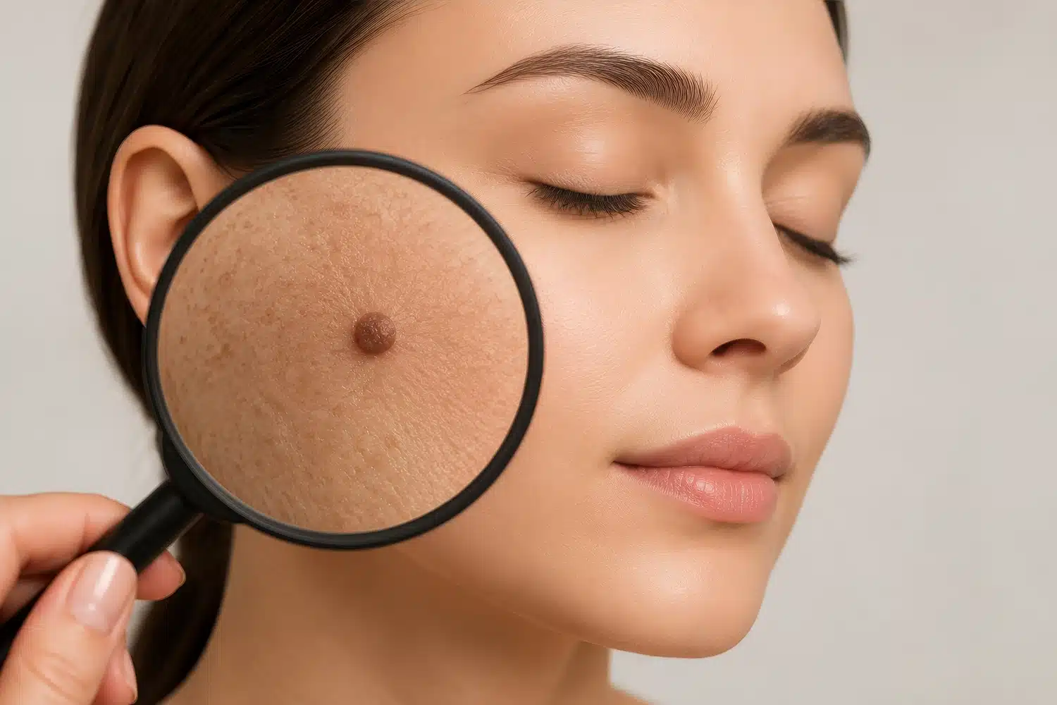
Appearance alone is not a reliable indicator of a mole's clinical significance, which is why a structured assessment is always the appropriate first step.
A mole may look entirely benign to the untrained eye and still carry microscopic features that a clinician needs to assess. Equally, a mole that looks irregular or dark may turn out to be a completely benign seborrhoeic keratosis (a harmless, non-cancerous skin growth).
A thorough pre-removal evaluation at my practice begins with a detailed patient history covering how long the mole has been present, whether it has changed, whether it bleeds or itches, and whether there is a personal or family history of melanoma.
I will also examine:
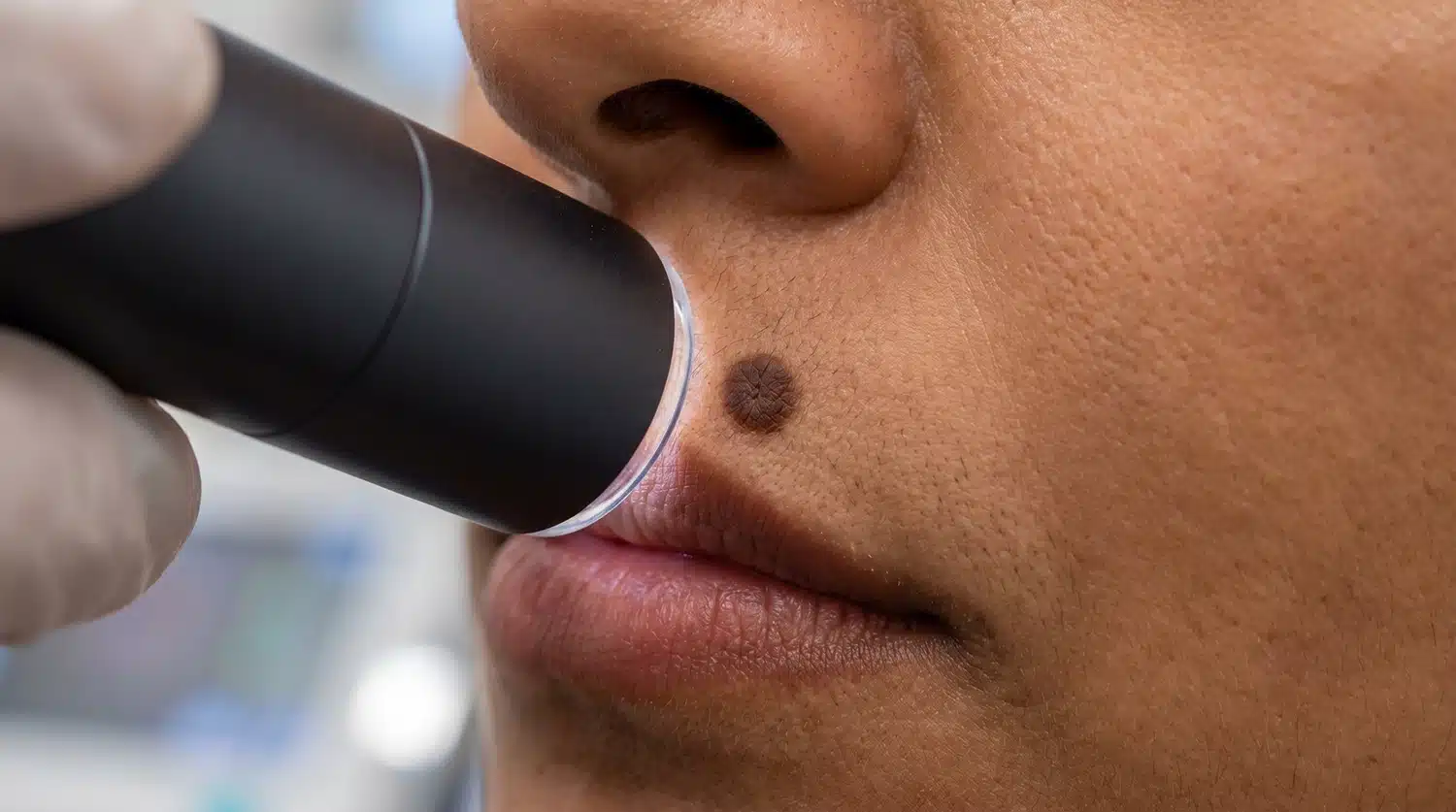
A dermoscope allows the clinician to examine the pigment network, blood vessel patterns, and the distribution of pigment dots and globules.
Two structural features are particularly significant: regression structures, where the skin appears to be replacing previously pigmented tissue, and blue-white veils, a diffuse area of bluish discolouration associated with certain types of melanoma.
Studies consistently demonstrate that dermoscopy improves diagnostic accuracy for melanoma detection compared to naked-eye examination alone, with sensitivity increasing from approximately 60% to over 85% in trained hands across published studies.
This is not a marginal improvement. It represents the difference between identifying a concerning mole and missing it.
In a benign mole, the pigment network is typically regular: lines of even thickness, equal spacing, and a pattern that fades gradually at the edges.
In a mole with atypical features, the network may be thickened, thinning abruptly, or terminating suddenly at the border.
At 1Aesthetics, I also examine dot and globule distribution, symmetry across two axes, and whether any structures fall outside the expected range for a benign lesion. A mole showing any of these atypical features warrants further clinical consideration. What dermoscopy identifies, or rules out, shapes every decision that follows.
Every assessment at 1Aesthetics includes dermoscopic examination before any recommendation is made. Book your mole assessment with me to understand what your mole's features mean before deciding on next steps.
This is one of the most clinically important principles in mole removal.
Laser ablation works by vaporising tissue. If that mole contained atypical or malignant cells, the evidence is destroyed along with it. There is no specimen to send to the laboratory, no pathology report, and no way to know whether the mole required further treatment.
Research confirms that laser therapy of pigmented lesions can delay melanoma diagnosis and may negatively affect outcomes when the disease has progressed further.
Surgical excision, by contrast, removes the mole intact and allows the entire specimen to be examined under a microscope. A pathologist can assess the cell architecture, confirm the excision margins, and recommend further management if necessary.
Laser microsurgery offers a middle ground in some cases, as it can preserve abnormal tissue for histological examination without removing the surrounding skin as excision biopsy would. However, because less tissue is removed, there is a greater chance of missing abnormal changes. In such cases, I may recommend surgical excision as the more thorough option where malignancy is a concern.
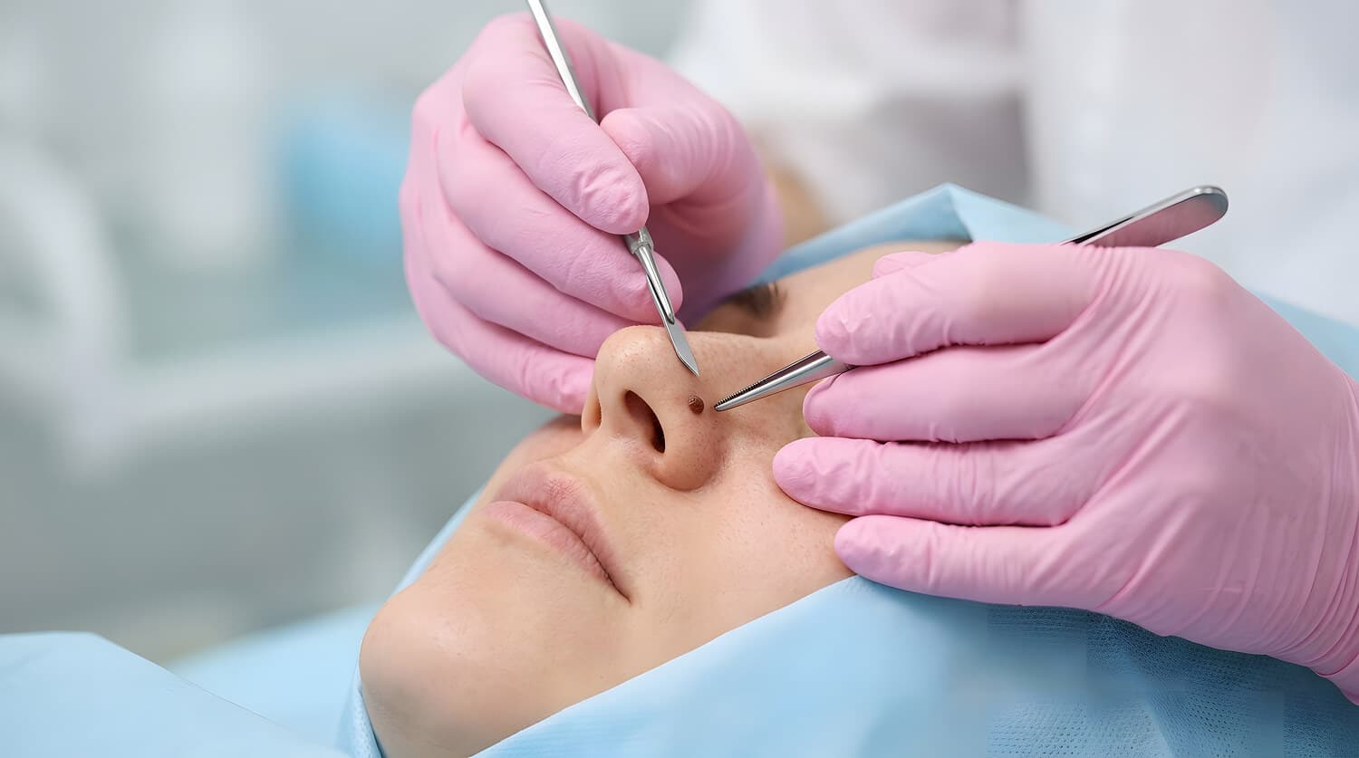
If a mole appears suspicious during dermoscopic examination, I may advise removing it and sending the tissue for laboratory analysis.
Any mole with features that raise clinical concern should be removed by surgical excision and sent for histopathological analysis, regardless of the patient's preference for a less invasive approach.
Not sure whether your mole is suitable for laser removal? Book an appointment with me at 1Aesthetics. I will examine the mole directly and recommend the approach that's clinically appropriate for your case.
For most patients, a mole found to be clinically benign simply needs to be removed in the most appropriate way. The choice of mole removal method depends on the mole's size, depth, location, whether it is raised or flat, and the patient's skin type and healing history.
| Mole type | Likely removal approach |
| Flat or superficial moles | Laser ablation or pigment laser treatment |
| Raised moles of moderate depth | Radiofrequency ablation or shave excision |
| Larger, deeper, or high-precision location moles | Surgical excision with suture closure |
At 1Aesthetics, no removal method is recommended, and no price is quoted without first examining the lesion directly.
Monthly skin self-examination is a reasonable practice for most patients. Examine your skin in good lighting, use a mirror for hard-to-see areas, and notice when something changes.
Annual skin checks with a doctor are recommended as a general guide for patients who have had an atypical mole removed, who have a significant sun exposure history, or who have more than 50 moles, in line with guidance on enhanced surveillance for atypical moles.
Certain signs should prompt a clinical review without delay:
These signs do not necessarily indicate melanoma, but they do indicate that a clinical assessment is needed promptly.
Whether your motivation for mole removal is cosmetic or clinical, the process begins in the same place: a thorough examination of the mole itself.
At 1Aesthetics, Dr Wan Chee Kwang carries out a comprehensive assessment before recommending any removal method. This includes dermoscopic examination, a review of your personal and family history, and a clear explanation of what the mole's features mean for your next steps.
Book an assessment at 1Aesthetics before making any decisions about mole removal.
Address:
1Aesthetics, Medical & Surgery
#14-90 The Central Tower 1
8 Eu Tong Sen Street
Singapore 059818
Mon-Fri 10 AM to 730 PM
Sat 10 AM to 6 PM
Phone / WhatsApp:
+65 66125173 / +65 84899962
Email:
[email protected]